Loading...

Archive

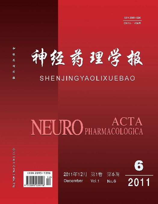
    26 December 2011, Volume 1 Issue 6 Previous Issue    Next Issue

    For Selected: Toggle Thumbnails
    Protective Effects of Emodin on the Memory of Mice Experiencing Cerebral Ischemia Reperfusion
    WANG Shu, ZHANG Li, XUE Gui-ping
    2011, 1 (6):  1-6. 
    Abstract ( 3228 )   PDF (1325KB) ( 2065 )  
    Objective: To study the protective effects of emodin on the memory of mice experiencing cerebral ischemia reperfusion and their underlying mechanisms. Methods: Using improved Himori method, cerebral ischemia reperfusion injury was produced in conscious mice by temporarily obstructing bilateral common carotid arteries. The protective effects of emodin (10.0, 1.0, 0.1 mg.kg-1,ip) on the memory of these mice were examined using the step-through test, shuttle box test,  antihypoxia test under normal pressure, test of acute cerebral hypoxia induced by decapitated mice and test of NaNO2 histotoxic anoxia,. Meanwhile, the superoxide dismutase (SOD) activity and glutathione peroxidase (GSH-PX) activity were also measured. Results: Emodin could attenuate the memory impairment induced by cerebral ischemia reperfusion injury, prolong the antihypoxic survival time, and increase the activities of GSH-PX and SOD. Conclusions: Emodin showed protective effects on the memory of mice with cerebral ischemia reperfusion injury. The possible mechanism may involve the enhancement of the activities of GSH-PX and SOD, which leads to the increased oxygen free radicals scavenging ability, enforced antihypoxic capacity and alleviation of cerebral ischemia damage.
    References | Related Articles | Metrics
    A Comparative Morphological Study on Early Stage Treatment of Rat Spinal Cord Contusion with Recombinant Human Erythropoietin and Methylprednisolone
    GU Bing, WANG Shuo-yu, LI Hua-nan, WANG Jun, ZHANG Shui-yin
    2011, 1 (6):  7-16. 
    Abstract ( 3422 )   PDF (16656KB) ( 1193 )  
    Objective:To comparatively study the morphological changes of the spinal cord in rats by treatment with recombinant human erythropoietin (rHuEPO) and methylprednisolone (MP) at an early stage after contusion injury. Methods:Spinal cord contusion model in rats was duplicated with a BenchmarkTM stereotaxic impactor. 30 min after the procedure, animals were administrated with 5000 UI·kg-1·BW rHuEPO (i.p.) or 30 mg·kg-1·BW MP (tail intravenous injection). Morphological changes were examined by using the Harris HE staining and Luxol fast blue staining methods in combination with the immunohistochemical NeuN staining and GFAP staining. Results: One and 3 days after the contusion procedure, the number of survived neurons, the residual myelination area and the reduction of astrocytes in the bilateral anterior horn of gray matter in and 3-5 mm around the the injury center were significantly higher in MP group than in rHuEPO group. However, 14 and 28 days after the contusion procedure, the cavity area of and surrounding the spinal cord injury site and the reduction of astrocytes were significantly larger in rHuEPO group than in MP group. In addition, the number of survival neurons and residual myelination area 3, 5 mm surrounding the injury site were significantly larger in rHuEPO group than in MP group (P<0.05). ConclusionMP (30 mg·kg-1·BW)treatment at the early stage after contusion damage (the first week) significantly improves the neuronal survival, reduces loss of the myelin sheath and inhibits the proliferation of astrocytes in rats with spinal cord injury. However, at a later stage (2-4 weeks after the injury) the effect of rHuEPO treatment was more significant.
    References | Related Articles | Metrics
    Effects of Silibinin on Depression-like and Anxiety-like Behaviors Induced by Sodium Butyrate in Aged Mice
    ZHANG Yan, WANG Jia-xing, LIU Na, BU Yu-jie, BAO Jin-feng
    2011, 1 (6):  17-21. 
    Abstract ( 3301 )   PDF (1258KB) ( 2210 )  
    Objective: To explore the behavioral dysfunctions induced by sodium butyrate in aged mice and the prophylactic effects of silibinin. Methods: Eight-month-old kunming mice were randomly divided into control group, sodium butyrate group and sodium butyrate plus Silibinin (0.1, 0.2 g·kg-1) group. The behaviors of aged mice were examined via the tail suspension test, open field test, elevated plus maze test and light/dark box test. Results: In the tail suspension test,the immobility time of sodium butyrate group was significantly increased compared with control group (P<0.05), while silibinin significantly antagonized the increase in immobility time induced by solidum butyrate (P<0.05). In the open-field test, the total travel distance was significantly decreased in solidum butyrate group as compared with control group (P<0.05) and silibinin treatment reversed such a decrease. In the elevated plus maze test, time spent in closed arms after sodium butyrate treatment in mice was significantly increased as compared with the control group (P<0.05), and there was a trend that silibinin blocked such an increase but failed to reach statistical significance.In light/dark box test, the residence time in the light box for the mice in sodium butyrate group was significantly decreased than for those in control group (P<0.05). Silibinin (0.2 g·kg-1) treated mice exhibited a significantly increased residence time of the light box compared with sodium butyrate group (P<0.05). Conclusion: Sodium butyrate can induce depression-like and anxiety-like behaviors. Silibinin can relieve those behavioral changes induced by sodium butyrate.
    References | Related Articles | Metrics
    A Method of Separating and Culturing Astrocytes from Rat Cerebral Cortex
    FAN Yue,YAN Yong,SHANG Ya-zhen
    2011, 1 (6):  22-27. 
    Abstract ( 2774 )   PDF (3194KB) ( 2617 )  
    Objective: To establish a method for rat cerebral cortical astrocyte culture, purification and identification. Methods: The cerebral cortex cells of newborn rats (1-3 days old) were cultured. The fibroblast cells and neurons were removed through differential adhesion and passage. Glial fibrillary acidic protein (GFAP), an astrocyte specific protein, was used for identifying astrocytes by immunohistochemical method. Results: The cultured cells showed typical morphological features of astrocyles and grew well. The third generation of the cells comprised > 95% astrocytes. Conclusions:The method is a simple and reliable in vitro culture for obtaining high purity astrocytes from newborn rat cerebral cortex.
    References | Related Articles | Metrics
    Animal Models of Alzheimer’s Disease and Pharmacotherapeutics
    JIN Miao, AN Hong-mei
    2011, 1 (6):  28-36. 
    Abstract ( 5575 )   PDF (1296KB) ( 3562 )  
    This paper reviewed recent progress in animal models of Alzheimer's disease (AD) and pharmacotherapy of AD. Herein, we summarized the different types of AD animal models based on the pathogenesis of AD, and concluded that there is a lack of full demonstration of the etiology and pathological changes of AD disease in animal models. We also summarized the current available medication treatments of AD. Aβ and Tau protein are still the research focus in the field of AD treatment. Chinese medicine also plays a certain role in the treatment of AD, and shows a good prospect.
    References | Related Articles | Metrics
    Progress in Understanding the Pathogenesis of Alzheimer’s Disease
    XUE Xiao-yan, GUO Xiao-hua, LI-Min, LUO Huan-min
    2011, 1 (6):  37-47. 
    Abstract ( 3497 )   PDF (1307KB) ( 3637 )  
    Although thepathogenesis of Alzheimer’s Disease (AD) is unclear, several hypotheses have been proposed, including the β-amyloid cascade theory, tau protein hypothesis, inflammation hypothesis and oxidative stress theory. This review summarized the widely-accepted new viewpoints on the pathogenesis of AD.
    References | Related Articles | Metrics
    Progress in Understanding the Relevance of Learning and Memory to Estrogen and Glu-NMDA Receptor Pathway
    ZHANG Xia-wei, ZHANG Dan-shen
    2011, 1 (6):  48-59. 
    Abstract ( 4025 )   PDF (1338KB) ( 3566 )  
    Besides maintaining the female secondary sexual characteristics, estrogen also plays important roles in the nervous system, especially in the brain: it can protect tissue from cerebral ischemia reperfusion injury and reduce infarct area by suppressing free radical formation, and can also regulate the release of excitatory amino acids. As an antioxidant, estrogen can protect the nervous system by regulating the body’s redox homeostasis. In addition, estrogen can play its neuroprotective role through estrogen receptors (ER). ER-α and ER-β, two typical ER subtypes that are mainly located in the nucleus, are both expressed in the cerebral cortex and hippocampus. It has been found that ER-α level in the cortex and hippocampus of ovariotomy rats is decreased accompanying decreased learning and memory. LTP, synaptic density and learning and memory are all affected in ER-β gene knock-out mice. NMDA receptor is critical in learning and memory and is coexpressed in hippocampus with estrogen receptors. Estrogen may activate ERK1/2 signal transduction pathway through membrane estrogen receptors, thus inducing the phosphorylation of the NR2B subunit of NMDA receptor to activate NMDA receptor. In addition, the synaptic plasticity can be affected by estrogen and its receptors, as well as by NMDA receptor.
    References | Related Articles | Metrics
    Recent Developments in the Study of Relationship between Wnt Signaling Pathway and Angiogenesis
    CHENG Hua, SUN Fang-ling, AI Hou-xi, ZHANG Li, JIANG Ying, WANG Wen
    2011, 1 (6):  60-63. 
    Abstract ( 3104 )   PDF (1184KB) ( 3083 )  
    There is a close relationship between Wnt signaling and angiogenesis, which plays a crucial role in the vascular endothelial cell proliferation, and in the remodeling, sprouting and maturation of blood vessels. Detailed investigation on the Wnt signaling pathway might reveal potential new targets for the treatment of neural diseases. This review focused on the relationship between Wnt signaling pathway and angiogenesis.
    References | Related Articles | Metrics